Anatomy!
Anatomy. The subject I dreaded before beginning at Warwick. So much to learn and so many difficult names! In my previous degree we had some anatomy but it wasn’t taught very well and therefore I remembered near to nothing when I started Medicine. However, even though I still find that it can be difficult, I have found it a lot less daunting than I first thought it would be.
We started pretty much straight away with anatomy, and that first lecture was a killer. Thankfully though that wasn’t the case for the rest of the year and I recovered! After I got used to the terminology and got my hands on a copy of Gray’s, things massively improved. The anatomy lectures were the best delivered lectures in my opinion and complicated topics were always covered again to help you process the information. We also had access to online recordings of the lectures, and extras for further help, so that meant we could go over the content ourselves if we needed to in our own time.
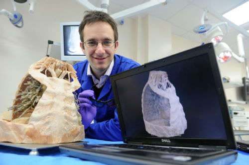
At the beginning of each module we were supplied with an anatomy workbook. Every week there would be questions to answer and pictures to be labelled, before a session in the surgical labs at University Hospitals Coventry and Warwickshire where we spent time with plastinated specimens and had smaller group teaching. Going through the workbooks before the session really helped to cement the content of that week’s lectures and made it easier to engage in the anatomy sessions at the hospital (they also made for a great revision tool). The sessions themselves were also great. I loved getting to see the specimens and begin to appreciate the relationship between different tissues.
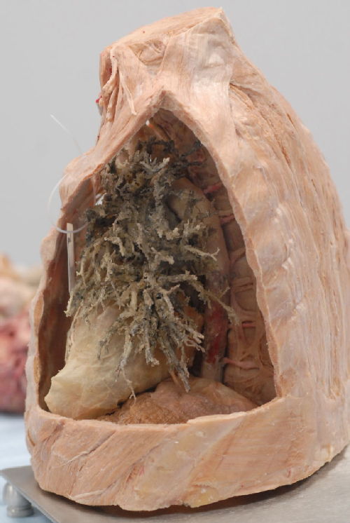
We also had access to 3D recordings of the specimens so that we could zoom in and appreciate a particular structure in more detail. At the end of the year we got to spend time with fresh tissue and this was fascinating. Having it at the end of the year meant we could name and identify a lot of structures that we probably never knew existed when we started! We also had revision stations at the end of each module with clinical cases based on the anatomy we had covered. This was a really helpful way to put everything together and understand how important being able to apply our anatomy teaching is.
During these anatomy sessions we also had seminars, where we would cover difficult aspects of a topic in more detail and at a slower pace, or ask questions about anything we had struggled with. I found these seminar sessions extremely useful. The repetition of key information through lectures, the workbooks and the seminars meant that things slowly made sense and were (hopefully) remembered when it came to exams!
We had radiology seminars during the anatomy sessions as well. It was a challenge, and still is, to be able to orientate and identify what different images were showing but these sessions were great at gradually introducing us to the world of x-rays, MRI and CT scans. We even had sessions where we could have a go at using ultrasound to look at each other’s abdomen and chest. This was great fun, albeit difficult, and we began to understand what the position of the probe meant we were actually seeing!
Having access to all of these different resources and teaching methods really helped with my learning and understanding of anatomy – and hopefully this will continue to be the case!
 Megan Earle
15 Oct 2014 11:13
|
Megan Earle
15 Oct 2014 11:13
| ![]() Tags: Anatomy Mbchb Rachel Warwick Wms
|
Tags: Anatomy Mbchb Rachel Warwick Wms
|  Comments (0)
|
Comments (0)
|  Report a problem
 Loading…
Add a comment
You are not allowed to comment on this entry as it has restricted commenting permissions.